Study Design. After anterior cervical discectomy, fusion was radiologically, biomechanically, and histologically assessed in a sheep spine fusion model.
Objective. To evaluate the efficacy of a platelet-rich plasma (PRP) application combined with a mineralized collagen matrix (MCM) as an alternative to autologous cancellous iliac crest bone grafts in a spine fusion model.
Summary of Background Data. PRP has the ability to stimulate bone and tissue healing. MCM is a recently developed osteoconductive material. Up to now, no comparative evaluation of PRP in combination with a MCM at the cervical spine has been performed in vivo.
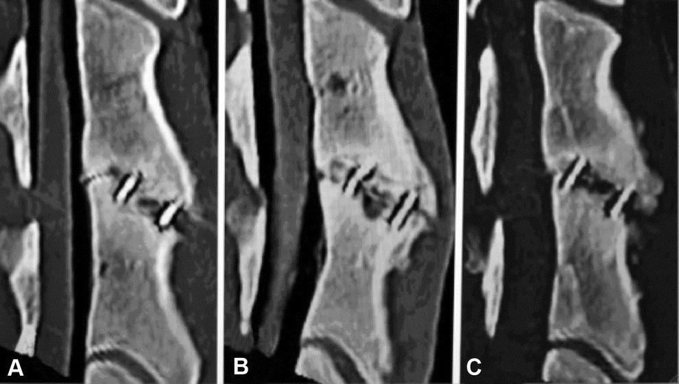
Methods. Twenty-four sheep (N = 8/group) underwent C3/4 discectomy and fusion: group 1, titanium cage filled with autologous cancellous iliac crest bone graft; group 2, titanium cage filled with MCM; and group 3, titanium cage filled with MCM and PRP. Radiographic evaluation was performed before surgery and after 1, 2, 4, 8, and 12 weeks, respectively. After 12 weeks, fusion sites were evaluated using functional radiographic views and quantitative computed tomographic scans to assess bone mineral density. Furthermore, histomorphologic and histomorphometrical analyses were performed to evaluate fusion.
Results. In comparison with the titanium cage group filled with autologous cancellous iliac crest bone grafts representing the control group, MCM-alone group showed a slightly lower fusion rate in the radiographic and the histomorphometrical analysis. The addition of PRP could not enhance this finding. There was no significant difference between MCM and MCM + PRP group in radiologic and histologic findings.
Conclusion. The MCM alone is not able to replace autologous bone grafts. Early activation of the platelets by calcium, which is released from mineralized collagen, could be the reason for the insufficient osteoinductive effect of PRP. In consequence, the combined application of mineralized collagen and PRP had no significant osteoinductive effect in this model.
