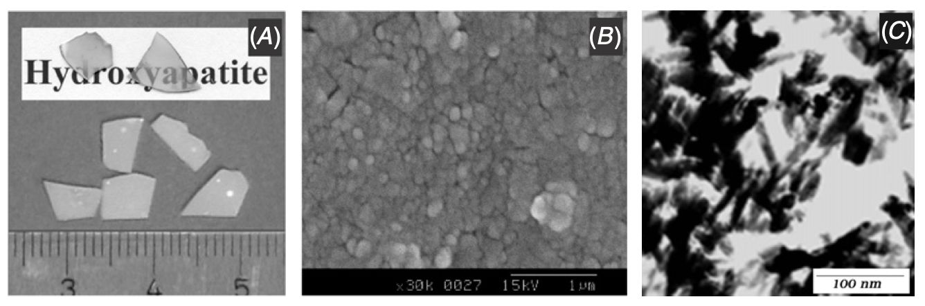
The light microscopic examination of cells directly on bioceramic materials in the transmission mode is impossible because many of these materials are opaque. In order to enable direct viewing of living cells and to perform time-lapse studies, nearly transparent bioceramic materials were developed. A dense and fine-grained transparent hydroxyapatite (tHA) was processed by a gel-casting route followed by low-temperature sintering (1000¡C). By virtue of its transparency, direct visualization of cellular events on this material is possible in transmitted light. In this study, the interaction of different bone cell types with the tHA ceramic was envisaged. Investigation of rat calvaria osteoblasts (RCO) cultured on tHA by means of transmission light microscopy indicated good cytocompatibility of tHA. Microscopic analysis of osteogenic-induced human bone marrow stromal cells (hBMSC) on tHA and quantitative analysis of their lactate dehydrogenase (LDH) activity at different time points of culture revealed favorable proliferation as well. An increase of the alkaline phosphatase (ALP) activity indicated the differentiation of osteogenic-induced hBMSC towards the osteoblastic lineage. In addition, the differentiation of human monocytes to osteoclast-like cells could also be demonstrated on tHA and was confirmed by fluorescent microscopy imaging of multinucleated cells on the transparent material.

The light microscopic examination of cells directly on bioceramic materials in the transmission mode is impossible because many of these materials are opaque. In order to enable direct viewing of living cells and to perform time-lapse studies, nearly transparent bioceramic materials were developed. A dense and fine-grained transparent hydroxyapatite (tHA) was processed by a gel-casting route followed by low-temperature sintering (1000¡C). By virtue of its transparency, direct visualization of cellular events on this material is possible in transmitted light. In this study, the interaction of different bone cell types with the tHA ceramic was envisaged. Investigation of rat calvaria osteoblasts (RCO) cultured on tHA by means of transmission light microscopy indicated good cytocompatibility of tHA. Microscopic analysis of osteogenic-induced human bone marrow stromal cells (hBMSC) on tHA and quantitative analysis of their lactate dehydrogenase (LDH) activity at different time points of culture revealed favorable proliferation as well. An increase of the alkaline phosphatase (ALP) activity indicated the differentiation of osteogenic-induced hBMSC towards the osteoblastic lineage. In addition, the differentiation of human monocytes to osteoclast-like cells could also be demonstrated on tHA and was confirmed by fluorescent microscopy imaging of multinucleated cells on the transparent material.