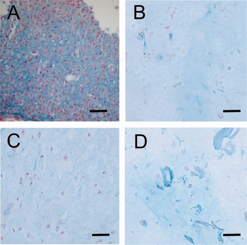
Although pellet culture and encapsulation of chondrocytes into gel-like biomaterials have lead to major advances in cartilage tissue engineering, a quantitative comparative characterization of cellular differentiation behavior during those cultivation procedures has not yet been performed. Our study therefore aimed at answering the following question: is the redifferentiation pathway of chondrocytes altered by slight changes in the type of alginate biomaterial (pure alginate, alginate-fibrin, alginate-chitosan) and how do the cells behave in comparison to biomaterial-free (pellet) three-dimensional culturing? Monolayer-expanded chondrocytes from healthy adult porcine knee joints were cultivated in alginate, alginate-chitosan, alginate-fibrin beads and as pellets up to 4 weeks. Quantitative PCR and Immunohistology were used to assess chondrogenic markers. Alginate-fibrin - encapsulated chondrocytes behaved almost like monolayer chondrocytes. Alginate- and alginate-chitosan encapsulation lead to a low chondrogenic marker gene expression. Although all 3D-cultured chondrocytes showed a considerable amount of Sox9 expression, only pellet cultivation lead to a sufficient Collagen II expression. This puts the usage of alginate-cultivated cartilage tissue engineering constructs under question. Fibrin addition is not beneficial for chondrogenic differentiation. Sox9 and Collagen II behave differently, depending upon the surrounding 3D-environment.

Although pellet culture and encapsulation of chondrocytes into gel-like biomaterials have lead to major advances in cartilage tissue engineering, a quantitative comparative characterization of cellular differentiation behavior during those cultivation procedures has not yet been performed. Our study therefore aimed at answering the following question: is the redifferentiation pathway of chondrocytes altered by slight changes in the type of alginate biomaterial (pure alginate, alginate-fibrin, alginate-chitosan) and how do the cells behave in comparison to biomaterial-free (pellet) three-dimensional culturing? Monolayer-expanded chondrocytes from healthy adult porcine knee joints were cultivated in alginate, alginate-chitosan, alginate-fibrin beads and as pellets up to 4 weeks. Quantitative PCR and Immunohistology were used to assess chondrogenic markers. Alginate-fibrin - encapsulated chondrocytes behaved almost like monolayer chondrocytes. Alginate- and alginate-chitosan encapsulation lead to a low chondrogenic marker gene expression. Although all 3D-cultured chondrocytes showed a considerable amount of Sox9 expression, only pellet cultivation lead to a sufficient Collagen II expression. This puts the usage of alginate-cultivated cartilage tissue engineering constructs under question. Fibrin addition is not beneficial for chondrogenic differentiation. Sox9 and Collagen II behave differently, depending upon the surrounding 3D-environment.
